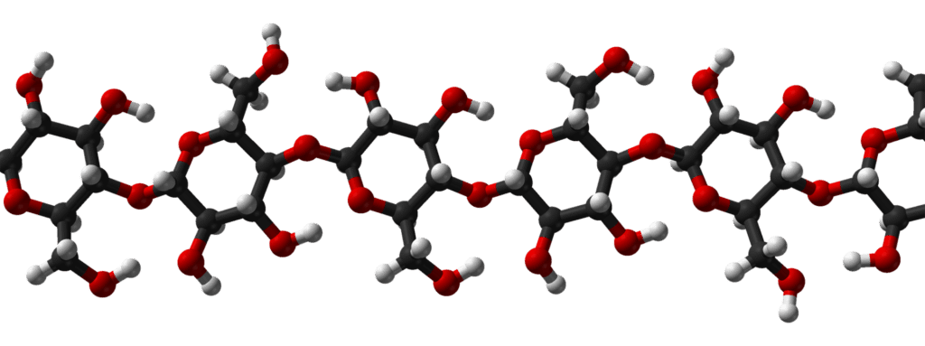
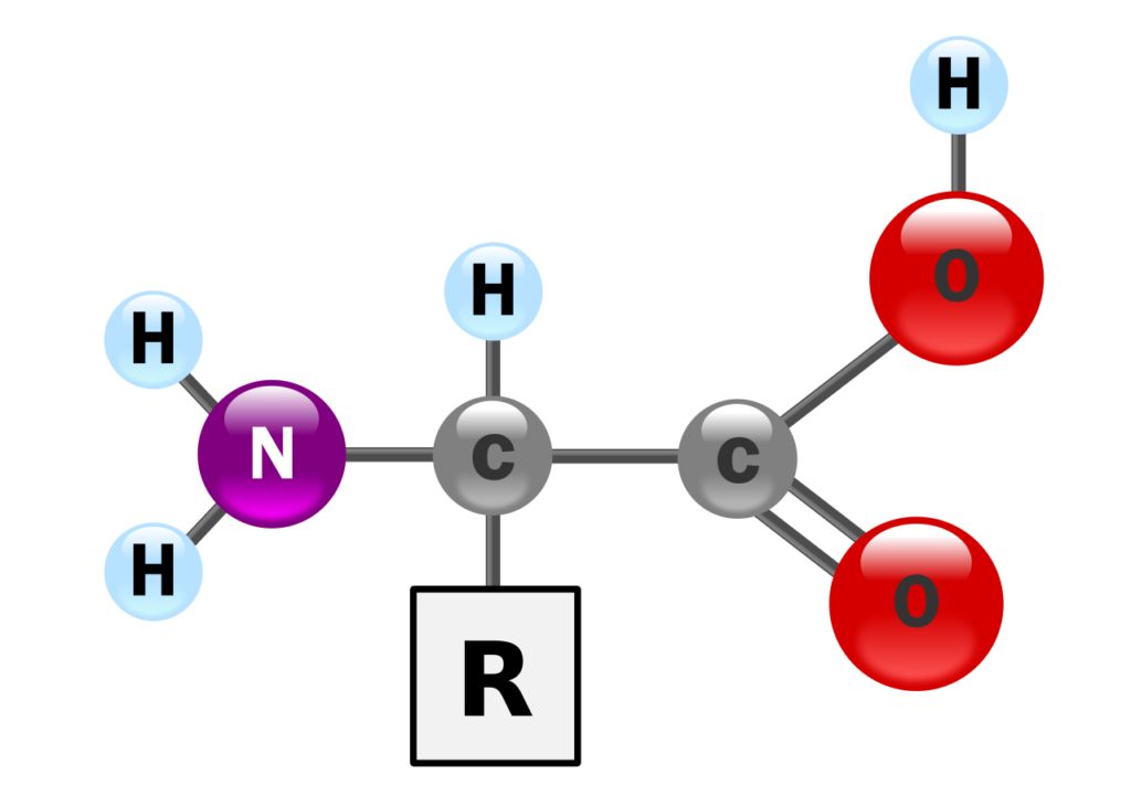
DNA is a long, linear polymer composed of four types of deoxyribonucleotides: adenine (A), cytosine (C), guanine (G), and thymine (T). These nucleotides are linked together by covalent phosphodiester bonds, which connect the 5′ carbon of one deoxyribose sugar to the 3′ carbon of the next.
The four bases attach to this repeating sugar-phosphate backbone, similar to beads of different colors on a string. The phosphodiester bonds give the DNA strand a specific polarity, with distinct 5′ and 3′ ends.
In 1953, American biologist James D. Watson and British physicist Francis H.C. Crick proposed a three-dimensional model for DNA structure. Their discovery earned Watson, Crick, and Maurice Wilkins the Nobel Prize in Medicine in 1962. The term “DNA” was introduced by Zaccharis.

Properties of DNA
- DNA serves as the hereditary material in nearly all prokaryotes and eukaryotes. It consists of two helical strands coiled around the same axis, and these helices can be either right-handed or left-handed.
- DNA is polar due to its highly charged phosphate-sugar backbone, which makes it soluble in water. However, in presence of salt and alcohol it is insoluble.
- DNA bases can absorb ultraviolet (UV) light at a wavelength of 260 nanometers, and the amount of UV absorption increases with the base sequence.
- When exposed to high temperatures, DNA denatures, as the heat breaks the hydrogen bonds between the base pairs, causing the two strands to separate. This temperature at which DNA denatures is called the melting temperature (Tm).
- The melting temperature is unique to each DNA sequence. Regions rich in cytosine-guanine (C-G) pairs have a higher melting temperature since more energy is required to break the three hydrogen bonds between these bases. In contrast, adenine-thymine (A-T) rich regions have a lower melting temperature because they are held together by only two hydrogen bonds.
- Upon cooling, the separated DNA strands can rejoin in a process known as renaturation.
Structure of DNA (Watson and Crick Model)
DNA can adopt different three-dimensional structures and is a flexible molecule. The Watson-Crick model is known as the B form of DNA, or B-DNA, which is the biologically significant form, as it is the most commonly found and naturally occurring form in living organisms. It is also the most stable under physiological conditions.

The important features of Watson – Crick Model or double helix model of DNA are as follows:
- The DNA molecule consists of two polynucleotide strands that coil around a common axis to form a right-handed double helix. The helices cannot be pulled apart but are separated during unwinding process, hence, called as plectonemic coil.
- These two strands run in opposite directions, making them antiparallel, with the 3′ end of one strand aligned with the 5′ end of the other i.e. if one helix of DNA molecule runs 5′ to 3′ direction, other runs in 3′ to 5′ direction, this is due to opposite orientation of sugar moieties in each helix.
- The sugar-phosphate backbones are located on the outside of the helix, while the purine and pyrimidine bases are positioned in the center.
- The strands are held together by hydrogen bonds between the purine and pyrimidine bases on opposite strands.
- Adenine (A) pairs with thymine (T) through two hydrogen bonds, while guanine (G) pairs with cytosine (C) through three hydrogen bonds. This specific pairing is called the base pairing rule, which makes the strands complementary.


- The sequence of bases along the polynucleotide chain varies, and this sequence encodes genetic information.
- DNA follows Chargaff’s rules, which state that the amounts of adenine (A) and thymine (T) are equal, as are the amounts of guanine (G) and cytosine (C). This means that the total purine content (A + G) equals the total pyrimidine content (C + T), and (A + C) is equal to (G + T). Additionally, the ratio of (A + T) to (G + C) is constant for a given species.
- The diameter of the DNA helix is 2 nm (20 Å), with adjacent bases spaced 0.34 nm (3.4 Å) apart along the axis.
- A full turn of the helix measures 3.4 nm (34 Å) and includes 10 base pairs per turn.
- The DNA helix has two grooves: a shallow minor groove with depth of 6 Å and depth of 7.5 Å and a deeper major groove having width of 12 Å and depth of 8.5 Å
Biological Functions of DNA
- Hereditary Material: DNA contains genetic information encoded in its nucleotide sequence, which is crucial for the synthesis of specific proteins or polypeptides and for passing this information to daughter cells or offspring.
- Autocatalytic Role of DNA: During the S-phase of the cell cycle, DNA replicates by using each strand of the double helix as a template to synthesize a complementary daughter strand.
- Heterocatalytic Role: In transcription, one strand of DNA acts as a template for RNA synthesis, fulfilling DNA’s heterocatalytic role.
- Variations: DNA undergoes recombination during meiosis and occasionally experiences mutations (changes in nucleotide sequences), leading to genetic variation in populations and driving evolution.
- Control of Cellular Functions: DNA oversees cellular metabolism, growth, and differentiation.
- DNA Fingerprinting: Also known as DNA typing or profiling, this technique is used to identify criminals, establish paternity, and verify immigration status.
- Recombinant DNA Technology (Genetic Engineering): This involves artificially cutting and recombining DNA from different organisms to create recombinant DNA, used to produce genetically modified organisms (GMOs), genetically modified foods (GMFs), vaccines, hormones, enzymes, and clones.


