Endoplasmic reticulum (ER) is the largest single membrane bound intracellular organelle found in eukaryotic cells that forms an extensive interconnected network of close and flattened membrane sacs or tube-like structures known as cisternae, mainly responsible for synthesis and transport of protein and lipid, metabolism of carbohydrate, compartmentalization of nucleus etc.
The ER occurs in most of the eukaryotic cells, but is lack in red blood cells and spermatozoa.
The enclosed compartment is called the lumen of ER. The membranes of the ER are contiguous with the outer nuclear membrane, even though their compositions can be different.
The ER is a dynamic structure that serves many roles in the cell including protein synthesis and transport, protein folding, lipid and steroid synthesis, lipid transfer and signaling to other organelles, compartmentalization of the nucleus, carbohydrate metabolism, detoxification of compounds, and calcium storage.
Early cytologists believed that cells contained a structural network or cytoskeleton, which was referred to by different names, such as Nissl substance, ergastoplasm, and basophilic bodies. In 1945, Porter, Claude, and Fullman observed a fine membranous network within the cytoplasm using an electron microscope. Keith Porter later named this structure the endoplasmic reticulum (ER) in 1953. Initially, the ER appeared to be restricted to the cell’s endoplasm, which influenced its naming.
Structure of Endoplasmic Reticulum
In eukaryotic cells, the endoplasmic reticulum is typically the largest membrane, forming an extensive network of interconnected sacs or channels. It constitutes about 30 to 60% of the cell’s total membrane.
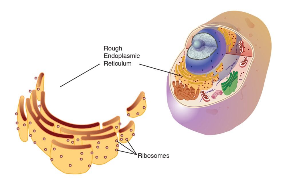
Ribosomes may or may not be attached to its outer membrane, leading to two types: rough (RER) and smooth endoplasmic reticulum (SER).
The rough ER, distinguished by ribosomes about 150 Å in diameter, is rich in protein and RNA. In contrast, the smooth ER lacks ribosomes and consists of three main structures: cisternae, tubules, and vesicles.
Cisternae
- Cisternae are flattened, unbranched, sac-like structures approximately 40-50 µm in diameter.
- They are arranged in parallel stacks, interconnected yet separated by cytosolic spaces.
- Ribosomes, which are small granular structures, may or may not be present on the surface of the cisternae.
Tubules
- Tubules are irregular, branching structures that create a network in conjunction with other components.
- They measure approximately 50-100 µm in diameter and are typically free of ribosomes.
Vesicles
- Vesicles are oval, vacuole-like structures with a diameter of around 25-500 µm, often found isolated in the cytoplasmic matrix and lacking ribosomes.
- The lumen of the ER contains a fluid known as the endoplasmic matrix, and all components of the ER are interconnected, allowing free communication among them.
The membrane surrounding the cisternae, tubules, and vesicles of the ER resembles the cell membrane, with a thickness of 50-60 Å. Composed of two phospholipid layers sandwiched between two layers of protein molecules, it has a relatively high protein-to-lipid ratio (Robertson, 1959).
The ER membrane is continuous with the cell membrane, Golgi membranes, and the outer nuclear envelope. Some cisternae open through pores in the cell membrane. Secretory granules, observed by Palade (1956), are present in the ER lumen, which serves as a pathway for secretory products.
Approximately 30-40 enzymes, supporting various synthetic activities, are associated with the ER, located on the cytoplasmic or luminal surfaces or both. The shape and size of membrane-bound ER spaces vary across different cell types.
Types of Endoplasmic Reticulum
On the basis of absence or presence of ribosomes, two kinds of Endoplasmic reticulum are found;
- Smooth Endoplasmic Reticulum
- Rough Endoplasmic Reticulum
Smooth Endoplasmic Reticulum (SER)
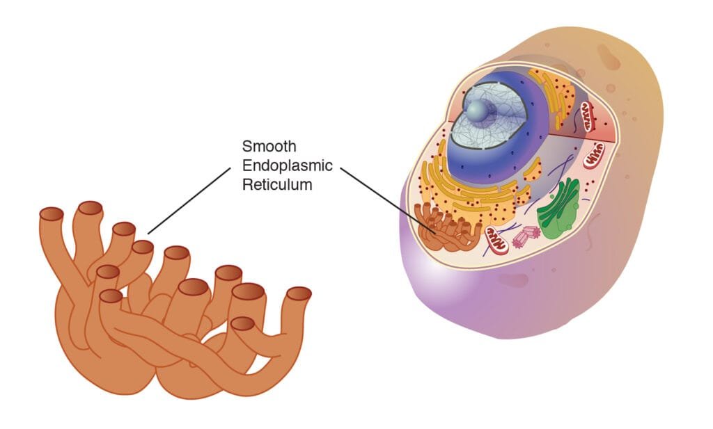
The absence of ribosomes on the ER’s surface gives it a smooth appearance, hence the name smooth or agranular ER.
It mainly exists in tubular forms, forming irregular lattices with a diameter of around 500-1000 Å.
SER is prominent in cells engaged in steroid or lipid synthesis (non-protein synthesis), such as those in adrenal and sebaceous glands or gonadal interstitial cells (Christensen and Fawcett, 1961).
Cells involved in carbohydrate metabolism (e.g., liver cells), impulse conduction (e.g., muscle cells), pigment production (e.g., retinal pigment cells), and electrolyte excretion (e.g., chloride cells in fish gills) also contain a high amount of SER.

Functions of Smooth Endoplasmic Reticulum(SER)
- Surface for Synthesis: The SER provides a surface for synthesizing fatty acids, phospholipids, glycolipids, steroids, and visual pigments.
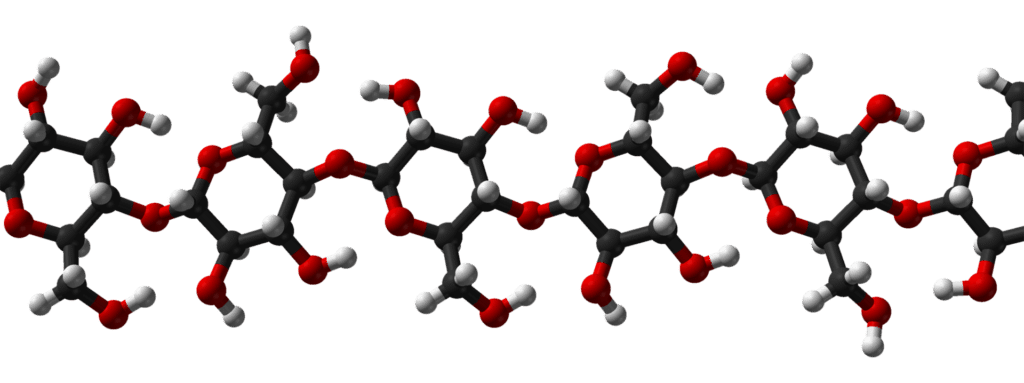
- Glycogen Metabolism: The SER contains enzymes essential for glycogen metabolism in liver cells, where glycogen granules are abundantly attached to the outer surface of SER membranes.
- Detoxification: SER enzymes in the liver facilitate detoxification, converting harmful substances like carcinogens and pesticides into harmless compounds for excretion.
- Formation of Organelles: The SER contributes to the formation of the Golgi apparatus, lysosomes, microbodies, and vacuoles.
- Transport Route: Proteins are transferred from the RER to the SER and then to the Golgi apparatus for further processing.
- Skeletal Muscle Contraction: In skeletal muscle cells, the sarcoplasmic reticulum releases Ca²⁺ ions to trigger contraction and absorbs them to promote relaxation.
- Fat Oxidation: Initial reactions for fat oxidation occur on the membranes of the SER.
Rough Endoplasmic Reticulum (RER)
The RER is distinguished by ribosomes on its surface, giving it a granular appearance.
It mainly takes the form of flattened cisternae around 400-500 Å wide. RER is prevalent in cells that actively synthesize proteins, such as enzyme-producing cells (e.g., pancreatic, plasma, and liver cells) or mucus-producing goblet cells.
In pancreatic exocrine cells, the RER forms reticular sheets and fenestrated cisternae in the cell’s basal region, with these cisternae measuring approximately 5-10 microns in length and grouped at diameters of 400-1000 Å.
In the cell’s apical region, the granular reticulum appears as vesicles. The membranes of granular and agranular ER are continuous where they connect.

Functions of Rough Endoplasmic Reticulum(RER)
- Surface for Ribosomes: The RER provides an extensive surface for ribosomes to attach.
- Surface for Synthesis: The RER offers a large surface area where ribosomes can efficiently carry out protein synthesis. Newly synthesized proteins may become incorporated into the ER membranes or enter the ER lumen. Those incorporated into the membrane eventually move from the ER through other organelles, such as the Golgi apparatus and secretory vesicles, to become part of the plasma membrane. Proteins entering the ER lumen are packaged for export.
- Packaging: Proteins in the ER lumen are processed and enclosed in spherical, membrane-bound vesicles that pinch off from the ER. These vesicles can have different outcomes: some stay in the cytoplasm as storage vesicles, while others move to the plasma membrane to release their contents via exocytosis. Some fuse with the Golgi apparatus for further protein processing, storage, or secretion from the cell.
- Smooth ER Formation: The RER gives rise to smooth ER when it loses its ribosomes.
- Formation of Nuclear Envelope: The RER helps form the nuclear envelope around daughter cells during cell division.
- Formation of Glycoproteins: The process of attaching sugars to proteins to create glycoproteins begins in the RER and is completed in the Golgi apparatus.
Overall Functions of Endoplasmic Reticulum(ER)
- The ER plays a crucial role in forming the skeletal framework by providing mechanical support through its membranous network, which helps hold various cell organelles in place.
- The membranes of the ER contain sites for various enzymes and cytochromes to carry out specific biochemical reactions.
- The ER increases the surface area available for cellular reactions, facilitating the rapid synthesis of biochemicals.
- The ER of one cell communicates with the ER of adjacent cells through structures known as desmotubules.
- The ER transmits information both from the external environment to the inside of the cell and between different organelles within the same cell.
- The ER acts as the cell’s circulating system, quickly transporting materials, such as proteins and carbohydrates, to other organelles, including lysosomes, Golgi apparatus, and the plasma membrane.
- During telophase, part of the nuclear envelope is formed by the ER, assisting in the creation of the nuclear membrane during cell division.
- The ER helps form vacuoles and provides membranes for the Golgi apparatus to produce vesicles and Golgian vacuoles.
- The ER supplies precursors to the Golgi apparatus for the complex formation and elaboration of biochemicals for both internal use and secretion.
- The ER is involved in the synthesis of proteins, glycogen, lipids, and steroids, such as cholesterol, testosterone, and progesterone.
Summary
Endoplasmic reticulum (ER) is the largest single membrane bound intracellular organelle found in eukaryotic cells that forms an extensive interconnected network of cisternae, mainly responsible for synthesis and transport of protein and lipid, compartmentalization of nucleus etc.
In 1945, Porter, Claude, and Fullman observed a fine membranous network within the cytoplasm using an electron microscope. Keith Porter later named this structure the endoplasmic reticulum (ER) in 1953.
On the basis of absence or presence of ribosomes, two kinds of ER are found in cells i.e smooth ER and Rough ER. The rough ER, distinguished by presence of ribosomes, rich in protein and RNA. In contrast, the smooth ER lacks ribosomes and consists of three main structures: cisternae, tubules, and vesicles.
Cisternae are flattened, unbranched, sac-like structures. Tubules are irregular, branching structures that create a network in conjunction with other components. And vesicles are oval, vacuole-like structures, often found isolated in the cytoplasmic matrix and lacking ribosomes.
ER plays a crucial role in forming the skeletal framework, providing surface area for cellular reactions, communicating with other cells, transporting components with in cell, synthesizing proteins, lipid, glycogen, hormones etc.


