Definition and Meaning
Plasma membrane, also known as the cell membrane is a selectively permeable membrane, present as the outermost, thin layer in animal cell and inner to cell wall in cell of plant, fungi, bacteria etc, which separates cells inner environment form external environment and also responsible for cell signaling, adhesion and protection of cell.
The plasma membrane, also known as the cell membrane or cytoplasmic membrane, outlines the cell’s boundaries and separates the cell’s internal environment from the external surroundings. This membrane, found in all cells, is a lipid bilayer embedded with proteins, sometimes called the plasmalemma.
It is selectively permeable, allowing only certain molecules and ions to pass through, thus controlling the movement of substances in and out of the cell.
The cell membrane plays crucial roles in processes such as cell signaling, where it transmits signals to other cells via receptors; ion conductivity, managing the exchange of charged ions across the membrane; and cell adhesion, enabling cells to interact and attach to neighboring cells.
Additionally, it provides a surface for various extracellular structures, including the cell wall, the glycocalyx (a carbohydrate-rich layer of glycoproteins and glycolipids), and the cytoskeleton, which is a dynamic network of protein filaments within the cell.
Karl W. Nägeli (1817–1891) demonstrated that the cell membrane is semipermeable and plays a role in osmotic and related phenomena in living cells. In 1855, Nägeli introduced the term “plasma membrane” to describe a firm protective layer that forms when cytoplasm flows out from a damaged cell, as protein-rich cell sap interacts with water.

Structure of Plasma Membrane
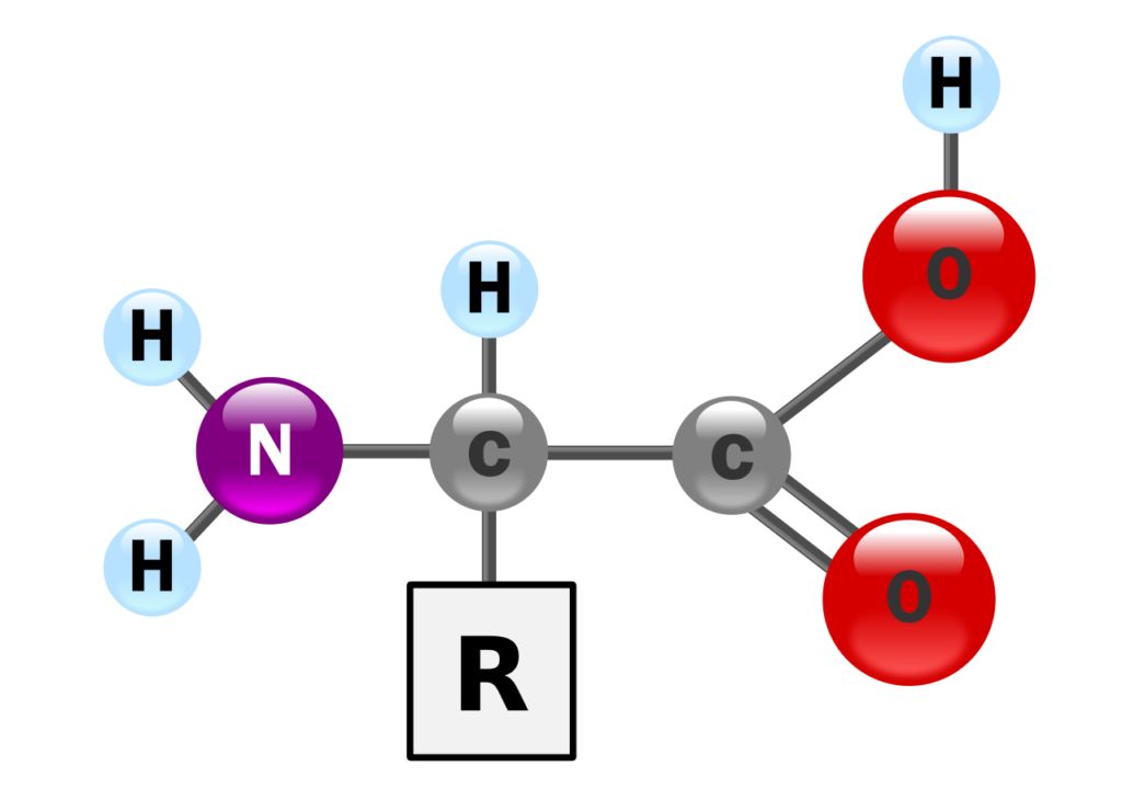
- The cell membrane contains structures that aid in the uptake of necessary solutes into the cell. Primarily composed of proteins and lipids, the membrane has a lipid layer that acts as a barrier, preventing mixing, while proteins enable the passage of polar substances and ions.
- Depending on their location and specific functions within the body, cell membranes can be made up of about 20 to 80% lipids, with the remainder being proteins.
- Proteins regulate and maintain the cell’s chemical environment and assist in the transport of molecules, while lipids add flexibility to the membrane.
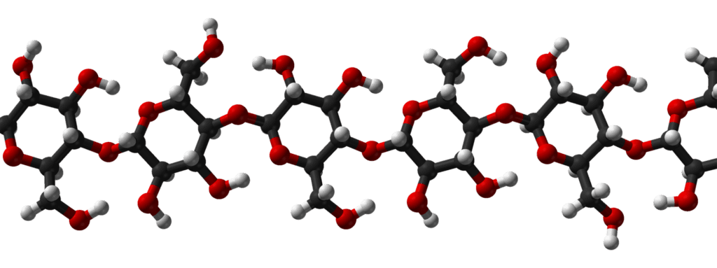
- Carbohydrates, making up about 5 to 10% of the membrane’s mass, are also present.
- They are attached to proteins as glycoproteins or to lipids as glycolipids, with an especially high concentration in the plasma membranes of eukaryotic cells.
Miceller Model of Plasma Membrane
- According to the mosaic model proposed by Hiller and Hoffman (1953), the plasma membrane is composed of globular subunits or micelles.
- When fatty acid molecules are fully surrounded by water, they can form structures called micelles, where the hydrophobic parts of the molecules face inward, away from the water, and the hydrophilic parts are exposed on the surface.
- These micelles can form small spheres or bimolecular layers, with tightly packed lipid molecules at the core and a shell of polar, hydrophilic groups.
- Each micelle has a diameter of about 40 to 70 Å. In the plasma membrane, the protein component forms a monolayer on each side of these lipid micelles, with the proteins represented as globular structures.
- Water-filled pores, approximately 4 Å in diameter, exist between the globular micelles and are bounded partly by the polar groups of micelles and partly by the polar groups of the proteins.
Fluid Mosaic Model of Plasma Membrane

- Scientists have used the fluid mosaic model to describe the structure of cell membranes.
- Various models were proposed to explain the structure and composition of the plasma membrane, but in 1972, Jonathan Singer and Garth Nicolson introduced the fluid mosaic model, which has become the most widely accepted.
- This model views membranes as semi-fluid structures with proteins embedded within a lipid bilayer, illustrating both the mosaic-like distribution of proteins across the membrane and the fluid mobility of lipids and proteins within the layer.
- The lipids, glycoproteins, and many intrinsic proteins within membranes are amphipathic, meaning they contain both polar and nonpolar regions.
- These molecules form a liquid crystal structure, with polar groups oriented toward the water phase and nonpolar groups positioned within the bilayer.
- The lipid bilayer provides a structural matrix that acts as a permeability barrier for the membrane.
- In membranes with a high lipid content, the bilayer is more extensive and only occasionally interrupted by protein molecules, whereas in protein-rich membranes, the bilayer is less prominent.
- The fluid mosaic model thus describes the molecular organization, composition, and ultrastructure of membranes.
- This arrangement allows enzymes and antigenic glycoproteins to display their active sites on the membrane’s outer surface.
- The membrane’s fluidity means that lipids and proteins have significant mobility within the bilayer.
- Lipid fluidity depends on the saturation level of hydrocarbon chains and ambient temperature; many membrane lipids are unsaturated, keeping the bilayer’s melting point below body temperature.
Functions of Plasma Membrane
- It preserves the cell’s individuality and shape.
- It keeps cell contents contained and separate from external substances.
- It shields the cell from injury.
- It regulates the movement of materials into and out of the cell, maintaining proper concentrations of molecules and ions to sustain cell life.
- The cell remains functional as long as the membrane can control what enters and exits.
- It forms distinct organelles within the cytoplasm.
- Its junctions hold cells together.
- Its inward folds aid in the intake of materials through endocytosis (pinocytosis and phagocytosis).
- Its outward folds (microvilli) increase surface area for nutrient absorption and form protective layers around cilia and flagella.
- Its receptor molecules allow information to enter the cell.
- Its oligosaccharide molecules assist in distinguishing between self and non-self.
- By controlling material and information flow, the plasma membrane enables metabolism.
- It allows the release of secretions and wastes through exocytosis.
- It regulates cellular interactions essential for tissue formation and immune defense. It aids certain cells in movement, forming pseudopodia in organisms like amoebas and leukocytes.
Summary
The plasma membrane, or cell membrane, is a selectively permeable barrier that forms the outermost, thin layer in animal cells and lies just inside the cell wall in plant, fungal, and bacterial cells. It separates the cell’s internal environment from the external surroundings and plays a key role in cell signaling, adhesion, and protection.
In 1855, Nägeli introduced the term “plasma membrane” to describe a firm protective layer that forms when cytoplasm flows out from a damaged cell, as protein-rich cell sap interacts with water.
Primarily composed of proteins and lipids, the membrane has a lipid layer that acts as a barrier, preventing mixing, while proteins enable the passage of polar substances and ions.
In 1972, Jonathan Singer and Garth Nicolson introduced the fluid mosaic model, which has become the most widely accepted to describe its structure. This model views membranes as semi-fluid structures with proteins embedded within a lipid bilayer, illustrating both the mosaic-like distribution of proteins across the membrane and the fluid mobility of lipids and proteins within the layer.
It is responsible for providing shape to cell, protecting cell, maintaining proper concentration of molecules or ions to sustain cell life, cell signaling, excreting waste materials out of the cell and regulating immune defense.


