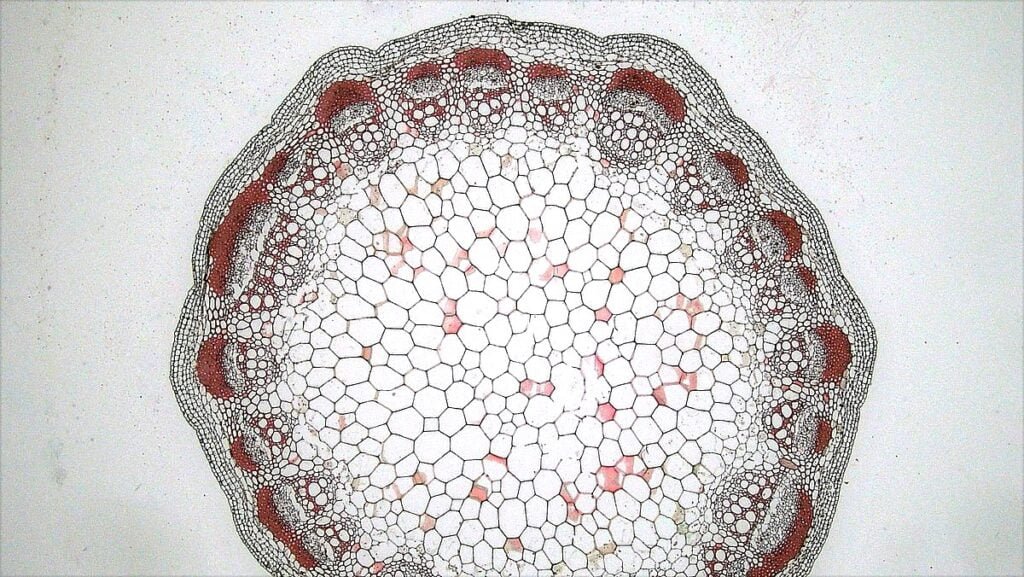
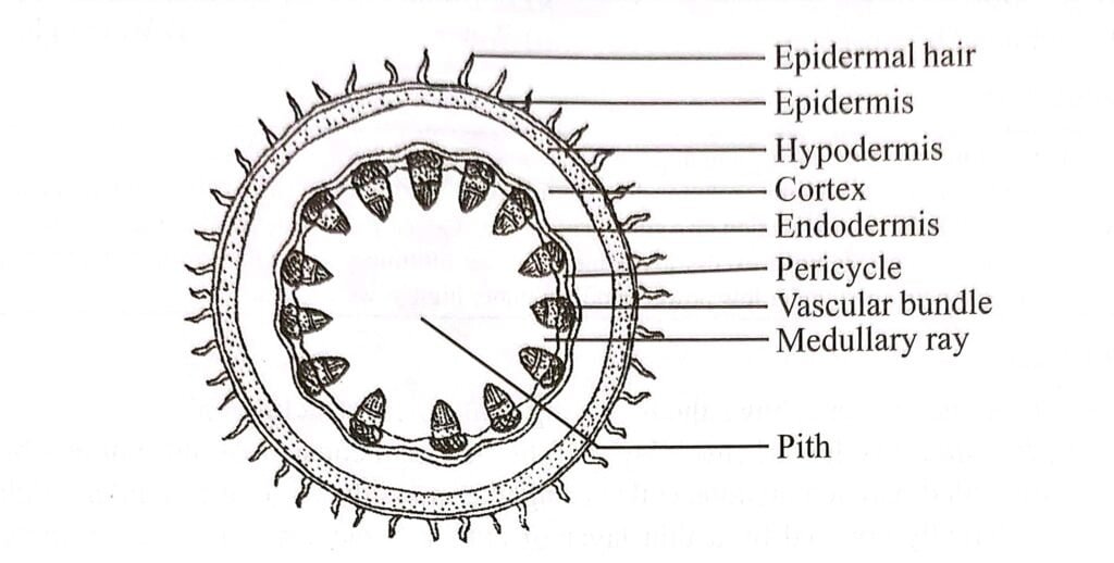
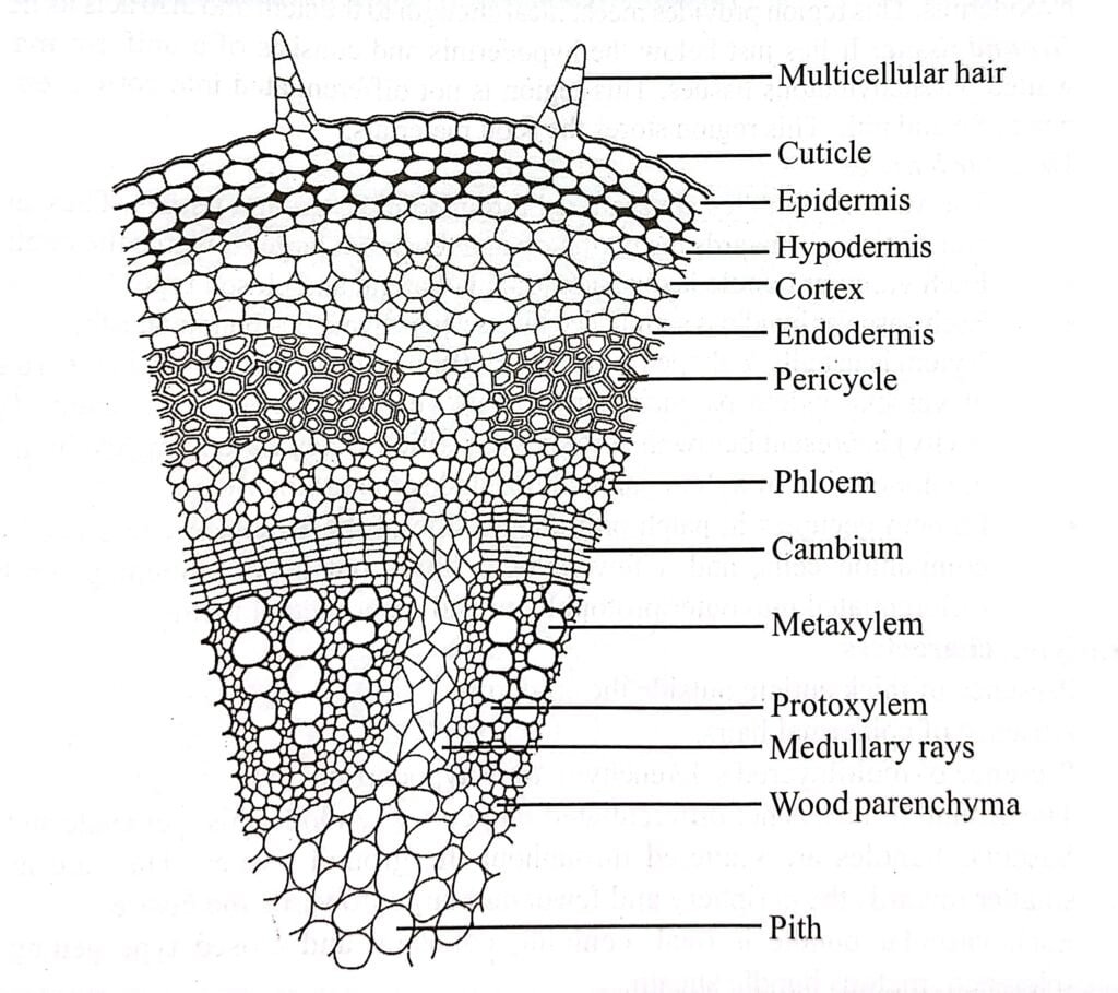
Internal structure of dicot stem, can be broadly defined by three structures; the outermost layer known as the epidermis, the central region consisting of vascular bundles, and the cortex that lies between the epidermis and the vascular bundles. However, these features can vary depending on the specific plant part. Anatomical structure in transverse section of typical dicot stem exhibits following features.
Epidermis
The outermost layer of the stem is known as the epidermis. It is composed of elongated, rectangular-barrel shaped parenchymatous cells that are tightly packed together.
These epidermal cells are transparent and do not contain chloroplasts. The outer walls of the cells are thick, convex, and cutinized, with a layer of cuticle present on the outer side. The inner walls of the epidermal cells are thin, while the radial walls gradually transition from thick towards the outer side to thin towards the inner side.
In the case, the epidermis bears unbranched multicellular hairs called trichomes, which are also covered by cuticle.
The epidermis serves various functions. It provides protection to the internal tissues of the stem, prevents the entry of harmful organisms, minimizes water loss through transpiration, allows for the exchange of gases through the stomata, and offers protection against excessive heating and sudden temperature changes, often aided by the presence of hairs.

Hypodermis
Beneath the epidermis, there is a multilayered collenchymatous tissue known as the hypodermis. The hypodermis typically consists of 3-4 layers of collenchyma cells located just below the epidermis.
These cells possess additional cellulose thickening in different regions of the cell walls. Collenchyma cells contain chloroplasts and enclose small intercellular spaces. Hypodermis is either absent or inconspicuous beneath the stomata
The hypodermis serves several functions, including providing mechanical strength and flexibility to the stem. It also acts as a storage site for food reserves and can perform photosynthesis when chloroplasts are present.
General Cortex
Beyond the hypodermis, there is a region known as the general cortex, which is composed of parenchymatous cells with intercellular spaces. The general cortex is a few to several cell layers thick.
The cells in the cortex can vary in shape, with some being angular and others being oval or rounded.
In young green stems, the outer cortical cells may contain chloroplasts, forming chlorenchyma, and contribute to food production through photosynthesis. However, the primary function of the cortex is to store food reserves.

Endodermis
This innermost layer of the cortex is known as the endodermis, and it acts as a boundary between the cortex and the vascular bundles. The endodermis consists of a wavy layer of cells that are one cell thick and have a barrel-shaped appearance.
Within the endodermal cells, conspicuous starch grains are often present, serving as a food reserve. This is why the endodermis of the stem is sometimes referred to as the “starch sheath.”
The general function of the endodermis is to store food reserves. It plays a role in storing starch and other nutrients within its cells.
Pericycle
The pericycle is found as patches of sclerenchyma located outside the phloem of each vascular bundle. It is a few cells thick and positioned inner to the endodermis and outer to the vascular strand.
The pericycle is a heterogeneous layer, composed of both parenchyma cells and sclerenchyma fibers, consisting of a completely sclerenchymatous wavy layer that is around 4-5 cells thick.
The sclerenchyma fibers form semicircular to semilunar patches known as bundle caps, which are located on the outer side of the vascular bundles. The parenchymatous pericycle is situated outside the medullary rays.
The sclerenchymatous pericycle provides mechanical strength to the young stem, offering support and rigidity. Meanwhile, the parenchymatous pericycle stores food reserves.
Vascular bundles
The vascular arrangement in the stem takes the form of an eustele, which refers to a ring of vascular bundles surrounding the central pith and located inner to the pericycle. These vascular bundles are present in a definite number and have an obtusely wedge-shaped appearance.
Each vascular bundle is composed of primary phloem on the outer side, primary xylem on the inner side, and a strip of cambium sandwiched between them. Both the phloem and xylem tissues are positioned on the same radius within the bundle.
These types of vascular bundles, characterized by the combination of phloem, xylem, and cambium, are known as conjoint, collateral, and open vascular bundles.
Xylem
- The xylem tissue is located towards the pith of the vascular bundles. It can be divided into two parts: the smaller protoxylem, which consists of narrow elements and is positioned closer to the pith, and the larger metaxylem, which consists of broader elements and is located on the outer side of the xylem. As a result, the xylem is considered endarch.
- The xylem tissue is composed of various cell types, including tracheids, vessels, xylem parenchyma, and xylem fibers.
- Among these, only the xylem parenchyma cells are living. They are smaller in size compared to the parenchyma cells found outside the vascular bundles. Xylem parenchyma cells serve the function of storing food reserves and aiding in the lateral conduction of sap within the xylem.
- Vessels are present in a few radial rows and have an angular outline. The vessels in the protoxylem region are smaller and possess annular or spiral thickenings. In contrast, the vessels of the metaxylem possess pitted thickenings.
- Tracheids are found between and around the radial rows of vessels, particularly in the metaxylem region. Xylem fibers are scattered amongst the tracheids.
- Both tracheids and vessels are involved in the conduction of water and minerals within the xylem. Additionally, the vessels, tracheids, and xylem fibers contribute to the mechanical strength of the stem, providing support and rigidity.

Phloem
- The phloem tissue is situated towards the pericycle on the outer side of the vascular bundle. It is composed of several components, including sieve tubes, companion cells, phloem parenchyma, and some phloem fibers.
- The sieve tubes are responsible for conducting organic food materials longitudinally within the plant. They are interconnected with companion cells and phloem parenchyma through pits. These connections facilitate the lateral flow of organic food within the phloem tissue.
- Additionally, companion cells play a crucial role in controlling the functions of the sieve tubes, ensuring efficient transport of nutrients.
- Phloem parenchyma cells are involved in storing various substances such as starch, proteins, and fats. These storage materials can be utilized when needed by the plant.
Cambium
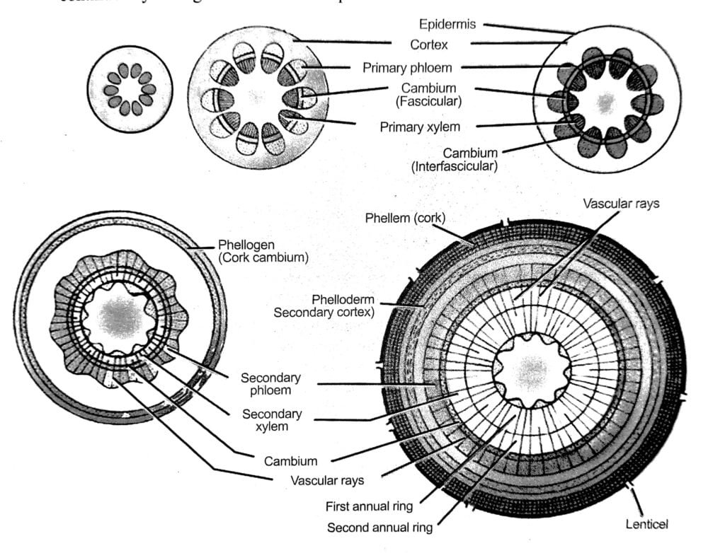
The cambium is a layer of meristematic tissue located between the xylem and phloem within the vascular bundle. It is derived from the pro-cambium, which is the initial meristematic tissue during plant development. The cambium appears as a thin strip of cells and is also known as the intrafascicular or fascicular cambium.
The cambial cells are thin-walled and elongated, taking on a fusiform shape that appears rectangular in transverse section. The primary function of the cambium is to contribute to the increase in stem girth through the production of secondary phloem towards the outer side and secondary xylem towards the inner side of the stem.
As the cambium cells divide and differentiate, they generate new cells that contribute to the growth of the stem in diameter. The production of secondary phloem and secondary xylem by the cambium helps in the overall thickening and structural support of the stem as the plant matures.
Medullary or Pith Rays
The medullary rays are radial strips of parenchyma cells that are located between adjacent vascular bundles in the stem. They serve as connections, linking the pith with the pericycle and cortex regions. The cells of the medullary rays are larger than those of the cortex and have a polygonal shape in cross-section. The intercellular spaces within the medullary rays are relatively small.
The ray cells of the medullary rays establish close contact with the conducting cells of both the phloem and xylem through pits, which are small openings in the cell walls. This intimate contact allows for the exchange and conduction of substances between the medullary rays and the vascular tissues.
The primary functions of the medullary rays include the radial conduction of food and water within the stem. They facilitate the movement of nutrients and water from the center of the stem (pith) to the outer regions (cortex) and vice versa. Additionally, the medullary rays play a role in the transport of gases, allowing for the exchange of gases between the pith and the cortex.
Pith or Medulla
The pith is the central region of the stem and is composed of polygonal, oval, or rounded parenchyma cells. These cells contain intercellular spaces, creating a porous structure within the pith. In certain dicot plants, the central part of the pith may disintegrate over time, resulting in the formation of a cavity known as the pith cavity.
The primary function of the pith cells is to store food reserves.


Pingback: Internal Structure of Monocot Stem - BioQuestOnline