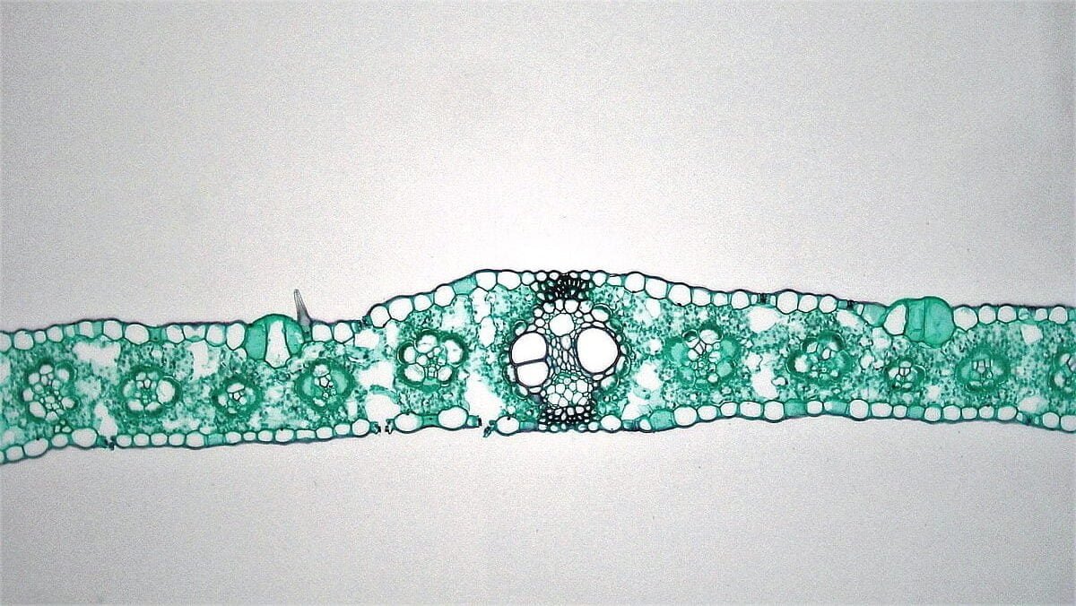
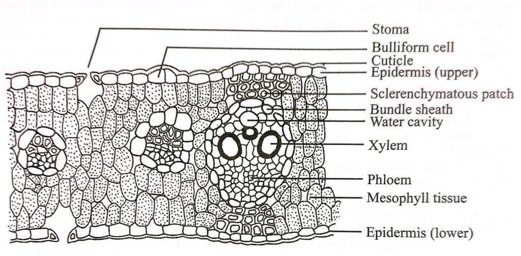
Internal structure of monocot leaf consist of epidermis (upper and lower), mesophyll tissue and vascular bundle.
In monocot leaves, the arrangement is isobilateral, meaning that both the upper and lower surfaces of the leaf are similar in appearance, align themselves parallel to main axis and direction of sunlight. They exhibit parallel venation.
Since, both sides receive a similar amount of sunlight. Consequently, both surfaces of the isobilateral leaf have a comparable level of greenness and possess a similar number of stomata.

Epidermis of Monocot (Isobilateral) Leaf
The leaf exhibits a single-layered epidermis on both its upper and lower surfaces called as upper epidermis and lower epidermis respectively. This epidermis is composed of closely packed barrel shaped parenchyma cells that are coated externally with a layer of cuticle (cuticularized).
Within the upper epidermis, there are specific cells known as bulliform cells or motor cells. These cells have a large size and thin cell walls and are capable of storing water when it is available. During periods of water scarcity, these bulliform cells lose water earlier than other cells, causing them to become flaccid. This leads to the rolling or curling of the leaf blade, which helps to minimize further water loss.
Both the upper and lower epidermal surfaces have a roughly equal number of stomata. Each stoma is surrounded by a pair of dumbbell-shaped guard cells.
The primary functions of the epidermis include protecting the internal tissues from microbial attacks, regulating water loss through transpiration, and facilitating gas exchange. The presence of the cuticle helps reduce the rate of transpiration, while the bulliform cells play a role in leaf protection by inducing leaf rolling and reducing transpiration.

Mesophyll tissue of Monocot (Isobilateral) leaf
The mesophyll tissue is situated between the upper and lower epidermis of the leaf. Unlike the differentiated palisade and spongy parenchyma found in dicot (dorsiventral) leaves, the mesophyll tissue in monocot leaves is relatively uniform or homogeneous in structure.
It consists of large, isodiametric oval or rounded cells with significant intercellular spaces or air cavities. The mesophyll tissue is made up of chlorenchymatous cells, meaning it contains a large number of chloroplasts with in it.
The primary function of mesophyll tissue is to carry out photosynthesis, the process by which plants convert sunlight into energy. And to store the prepared food material temporarily.
Vascular bundle of Monocot (Isobilateral) leaf
The mesophyll tissue contains numerous closely packed vascular bundles, ranging from small to large, which run parallel to each other, usually of smaller sized except at the midrib region.
Apart from the midrib, all the vascular bundles are embedded within the mesophyll tissue.
Each vascular bundle consists of phloem towards the lower epidermis and xylem towards the upper epidermis.
The vascular bundles are classified as conjoint, collateral, and closed type.

Each vascular bundle is surrounded by a single layer of compactly arranged parenchyma cells called the bundle sheath or border parenchyma. The midrib region, distinct from the rest of the mesophyll tissue, has deposits of sclerenchyma towards the upper and lower epidermis.
- Phloem: The phloem tissue within the vascular bundles is comprised of sieve tubes and companion cells. The sieve tubes conduct organic food, while the companion cells regulate the functions of the sieve tubes.
- Xylem: The xylem tissue contains vessels, tracheids, and xylem parenchyma cells. In small vascular bundles, the xylem is densely packed. Both vessels and tracheids are responsible for conducting water and mineral salts, as well as providing mechanical support to the leaves.


