Introduction
Digestive system of human is the group of organs responsible for ingestion, secretion, mixing & propulsion, breaking down (digestion), absorption and egestion of undigested food material. Digestive system of human consist of alimentary canal (mouth, pharynx, esophagus, stomach, small intestine, and large intestine) and accessory digestive organs (teeth, tongue, salivary glands, liver, gallbladder, and pancreas).
The digestive system begins with the mouth, which leads to a muscular tube called the pharynx. The pharynx continues into a long, tube-like structure known as the oesophagus, or food pipe. The oesophagus opens into the stomach, a relatively large chamber. The stomach is followed by the small intestine, which is divided into three parts: the duodenum (the beginning), the jejunum (the middle), and the ileum (the end). The small intestine then transitions into the final section of the alimentary canal, the large intestine. The large intestine is followed by the rectum, which ends in the distal opening called the anus.
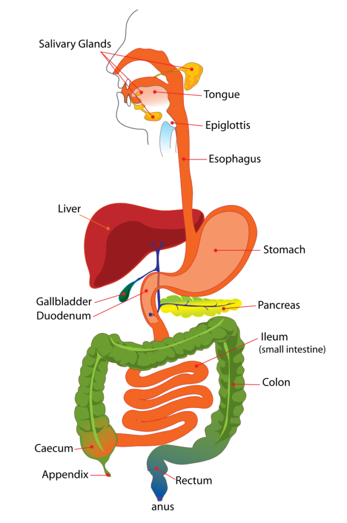
[Image Source: Mariana Ruiz ]
Over all process, digestive system of human perform;
- Ingestion: It involves taking foods and liquids into the mouth (eating).
- Secretion: Each day, cells within the walls of the GI tract and accessory digestive organs secrete a total of about 7 liters of water, acid, buffers, and enzymes into the lumen (interior space) of the tract.
- Mixing and propulsion: Alternating contractions and relaxations of smooth muscle in the walls of the GI tract mix food and secretions and move them toward the anus by a process called motility.
- Digestion: Mechanical and chemical processes break down ingested food into small molecules. mechanical digestion the teeth cut and grind food before it is swallowed, and then smooth muscles of the stomach and small intestine churn the food to further assist the process. As a result, food molecules become dissolved and thoroughly mixed with digestive enzymes while in chemical digestion, digestive enzymes catalyzes metabolic reactions which splits larger molecules in smaller ones which undergo absorption.
- Absorption: The entrance of ingested and secreted fluids, ions, and the products of digestion into the epithelial cells lining the lumen of the GI tract is called absorption. The absorbed substances pass into blood or lymph and circulate to cells throughout the body.
- Egestion: Wastes, indigestible substances, bacteria, cells sloughed from the lining of the GI tract, and digested materials that were not absorbed in their journey through the digestive tract leave the body through the anus in a form of feces or stool, called defecation or egestion..
Alimentary canal of Human
The gastrointestinal (GI) tract, or alimentary canal, is a continuous tube that extends from the mouth to the anus. Its about 5–7 meters in a living person. It consist of following parts;
- Mouth
- Pharynx
- Oesophagus
- Stomach
- Small intestine
- Large intestine
- Anal canal and Anus
The accessory digestive glands includes;
- Salivary glands
- Liver
- Pancreas
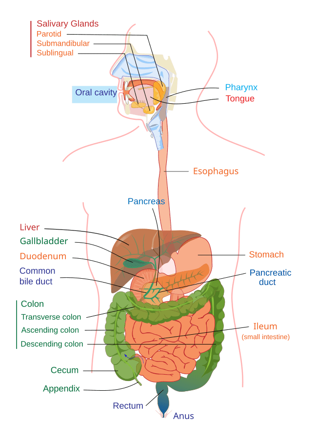
[Image Source: Mariana Ruiz]
Mouth
The mouth, also referred to as the oral or buccal cavity, is formed by the cheeks, palates, tongue and teeth.
Cheeks
- The cheeks form the sides of the oral cavity, with an outer layer of skin and an inner layer of mucous membrane. The front parts of the cheeks meet the lips, which are fleshy folds that surround the mouth’s opening.
- Each lip’s inner surface is connected to the gums by a midline fold of mucous membrane called the labial frenulum.
- The oral vestibule is the space between the cheeks and lips on the outside, and the gums and teeth on the inside. The oral cavity proper is the area extending from the gums and teeth to the fauces, which is the opening between the oral cavity and the oropharynx (throat).
Palate
- The palate is a partition that separates the oral cavity from the nasal cavity, forming the roof of the mouth.
- The hard palate is the front part of the roof of the mouth and creates a bony barrier between the oral and nasal cavities.
- The soft palate is the back part of the roof of the mouth, consisting of an arch-shaped muscular partition covered by a mucous membrane, separating the oropharynx from the nasopharynx.
- A fingerlike muscular structure called the uvula hangs from the free edge of the soft palate. When swallowing, the soft palate and uvula move upwards to close off the nasopharynx, preventing food and liquids from entering the nasal cavity.
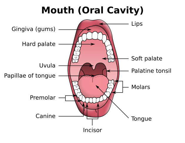
[Image Source: TilmannR, Wikimedia ]
Tongue
- The tongue, an accessory digestive organ made of skeletal muscle and covered with a mucous membrane, forms the floor of the oral cavity.
- The dorsal and lateral surfaces of the tongue are covered with papillae, which are projections of the lamina propria. Many papillae contain taste buds, the receptors for taste. Some papillae do not have taste buds but contain touch receptors, increasing friction between the tongue and food, which helps the tongue move food around in the oral cavity.
- The tongue’s lingual glands secrete both mucus and a watery serous fluid containing the enzyme lingual lipase, which breaks down triglycerides into simpler fatty acids and diglycerides.
Teeth
Teeth are accessory digestive organs located in the sockets of the alveolar processes of the mandible and maxillae, which are covered by the gingivae, or gums.
Each tooth has three main external regions: the crown, root, and neck.

- The crown is the visible part above the gum line, while one to three roots are embedded in the socket. The neck is the narrow junction between the crown and root near the gum line. Internally, dentin, a calcified connective tissue, forms most of the tooth and gives it shape and rigidity. Dentin is harder than bone due to hydroxyapatite and is covered by enamel, composed of calcium phosphate and calcium carbonate, which protects the tooth from the wear and tear of chewing. The pulp cavity, an enlarged space within the crown, is filled with pulp.
Humans consist of two sets of teeth i.e deciduous and permanent.

- Deciduous teeth, also known as primary, milk, or baby teeth, begin to erupt around 6 months of age, with about two teeth appearing each month until all 20 are present. These include 2 incisors, 1 canine, and 2 molars in each half of the upper and lower jaws. Deciduous teeth are typically lost between ages 6 and 12 and are replaced by permanent (secondary) teeth.
- The permanent dentition consists of 32 teeth: 2 incisors, 1 canine, 2 premolars, and 3 molars in each half of the upper and lower jaws. These teeth are responsible for biting, tearing, and grinding food before swallowing.
Pharynx
When food is swallowed, it moves from the mouth into the pharynx (throat), a funnel-shaped tube extending from the internal nares to the esophagus at the back and to the larynx at the front.
The pharynx is made of skeletal muscle and lined with a mucous membrane, and it is divided into three parts: the nasopharynx, oropharynx, and laryngopharynx.
The nasopharynx is involved only in respiration, whereas the oropharynx and laryngopharynx have both digestive and respiratory functions. Swallowed food passes from the mouth into the oropharynx and laryngopharynx, and the muscular contractions in these areas help move the food into the esophagus and then into the stomach.
Oesophagus
It is a collapsible muscular tube, about 25 cm long and 2 cm in diameter, lies posterior to the trachea. It begins at the inferior end of the laryngopharynx i.e cricopharyngeal sphincter passes through the inferior aspect of the neck, pierces the diaphragm through esophageal hiatus, and ends in the superior portion of the stomach guarded by cardiac sphcinter.
Oesophagus is responsible for passing food and liquids from the mouth down to the stomach by rhythmic contraction and relaxation of muscle, peristalsis. Food passes to stomach in the form of bolus.
Stomach
Stomach is wide J-shaped distendible, hollow muscular pouch like structure of about 30 cm long and 15 cm wide which is connected to the oesophagus at its upper end and to the duodenum at the lower end. It lies obliquely on the left side in the upper part of abdomen just below the diaphragm.
It has two curvatures. The right inner curve is called lesser curvature and the left outer curve is called greater curvature. Stomach is fixed to the body cavity by peritoneum called lesser and greater omentum.
Stomach consist of four parts:
- Cardiac part
- Fundus part
- Body part
- Pyloric part
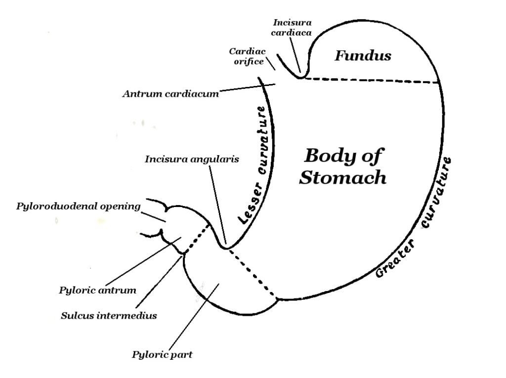
[Image Source: Gray, Henry, Public domain, via Wikimedia Commons]
- Cardiac part: It is the left narrow conical upper part of stomach into which oesophagus opens. It has opening called cardiac aperture guarded with cardiac sphincter. It checks the regurgitation of food. Cardiac tubular glands are found in the mucosa of cardiac part which secrete soluble mucus.
- Fundus part: It is the small dome like proximal part of stomach. It contains gas or air It also contains main gastric glands in mucosa layer.
- Body part: It is the middle main region of stomach. It contains infoldings called gastric rugae in mucosa and submucosa. It contains main gastric glands in mucosa layer.
- Pyloric part: It is the right narrow and tubular lower distal part. It has wider pyloric antrum and narrow pyloric canal. The pyloric canal has pyloric aperture guarded with pyloric sphincter. It opens into duodenum of small intestine. Pyloric glands are found in the antrum region of pylorus. These glands contain mucous cells and G-cells which secrete soluble mucus and hormone gastrin respectively.
Inner surface of stomach has numerous folds, called gastric rugae which makes the stomach distensible and can hold about 2-3 liter of food and water.
Main function of stomach is to storage of food (for 3-6 hrs. according to the nature of food), mechanical churning (mixing) of food, partial digestion of food, secretion gastric juice, hormone gastrin and a protein Castle’s intrinsic factor and absorption in small amount.
Small Intestine
Small intestine is a narrow and convoluted muscular tubular structure of about 6 m long and is the longest part of alimentary canal. It extends from the pylorus of stomach to caecum. It is fixed to the body wall by mesentery remain surrounded by the large intestine.
Small intestine consist of three parts;
- Duodenum
- Jejunum
- Ileum

[Image Source: National Caner Institute , Public Domain]
- Duodenum: It is the first and shortest part, about 25 cm long. It follows the stomach and curves around the head of pancreas. It is C-shaped. It receives the hepatopancreatic ampulla of hepatopancreatic duct. The villi are greatest and most numerous present in the mucosa of duodenum. It consist of crypts of Lieberkuhn and Brunner’s gland in the mucosa layer.
- Jejunum: It is the middle part of small intestine with abundant tongue like villi in the mucosa layer. It is about 2.5 m long. It has thicker and vascular wall than the ileum.
- lleum: It is lower part and the longest part of small intestine, about 3.5 m long opens into caecum of large intestine through ileocaecal aperture guarded with ileocaecal sphincter. It has finger like villi. lleum has white patches of tissues, called Payer’s patches.
The inner surface of small intestine has circular folds, called plica circulares. Mucosa is raised into numerous microscopic projections called villi. The free surface of the cells covering villi has microvilli, The pas circulares, villi and microvilli greatly increase the absorptive surface area of small intestine for absorption of food.
Small intestine serves the function of mixing chyme with intestinal juice, Secretion of intestinal juice and intestinal hormones, chemical digestion and absorption of digested food.
Large Intestine
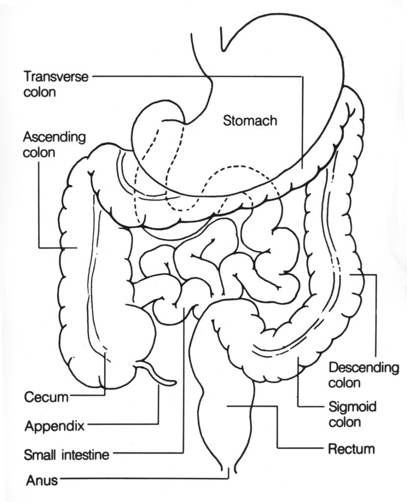
Large intestine is wide muscular tube of about 1.5 m long and 6 cm in diameter, arranged around the mass of small intestine in the form of question mark (?). It extends from the ileum to the anus and lacks villi and microvilli.
Large intestine consist of three main parts;
- Caecum
- Colon
- Rectum
[Image Source: National Cancer Institute : Public Domain]
- Caecum: It is the first part of large intestine. It is a small, blind pouch which receives ileocaecal aperture through which ileum opens. The ileocaecal aperture in guarded with ileocaecal sphincter which prevents backflow of intestinal contents. It has a worm like projection, called vermiform appendix which is about 8cm long. The inflammation of appendix is called appendicitis.
- Colon: It forms the major part of large intestine, li extends from caecum and consists of four parts ascending, transverse, descending and sigmoid colon. Ascending colon passes upward on the right side of abdomen. Transverse colon bends to the left and runs across the abdominal cavity. Descending colon extends downward on the left side of abdomen. Sigmoid colon turns to the right and joins rectum. The bend between the ascending and transverse colon is called hepatic (right colic) flexure and between the transverse and descending colon is called splenic (left colic) flexure. Mucous membrane of colon is smooth without villi. It gives colon a characteristic caterpillar appearance. The mucosa layer contains crescentic-shaped folds and crypts of Lieberkuhn.
- Rectum: It follows the colon. It is about 15-20 cm long. It is like a small dilated sac. It has longitudinal folds and large blood vessels. It leads through 4 cm long anal canal to anus.
Main functions of large intestine is to reabsorption of water and electrolytes, temporary storage and elimination of faeces, synthesizes certain vitamins (folic acid, vitaminB12, vitamin K) and excretion of heavy metals like mercury, lead, bismuth and arsenic.
Anal Canal and Anus
- Anal canal: It is 4 cm long and leads from rectum to the anus. It is lined with mucous membrane and the lower part with skin. The mucous membrane contains six to twelve longitudinal folds called anal columns.
- Anus: It lies at the base of anal canal which opens in the abdomen in a fold between the nates. It is guarded by two anal sphincters- Internal and external. Internal anal sphincter is made up of involuntary and external anal sphincter is made up of voluntary muscle fibers. It helps in egestion.
