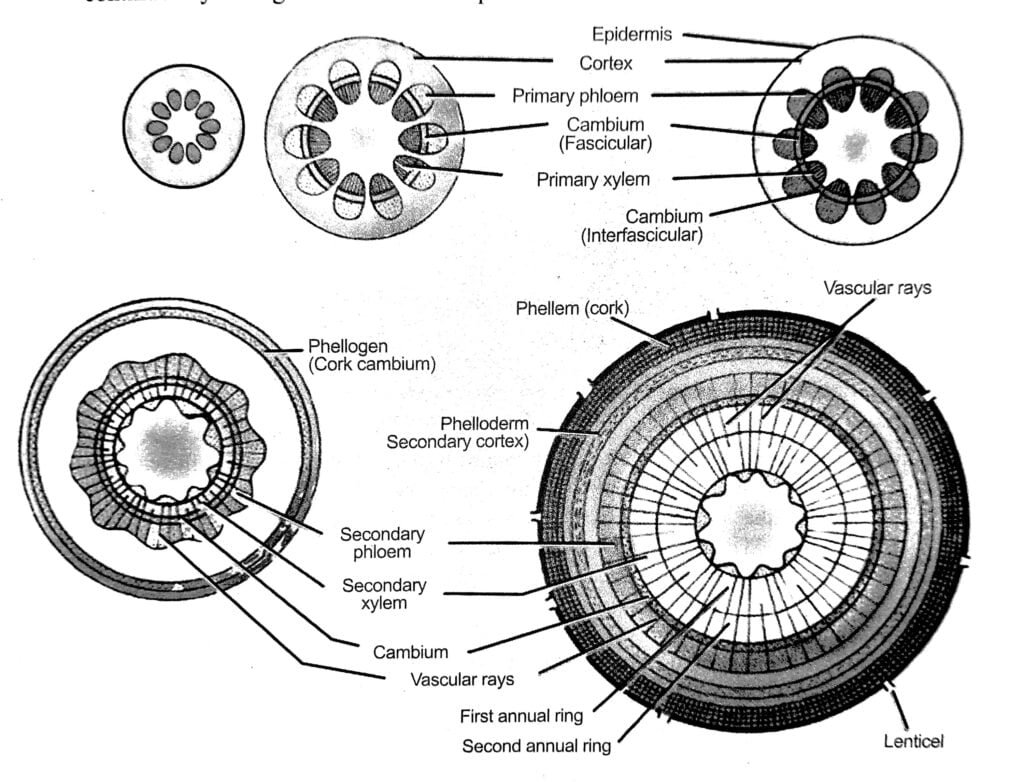
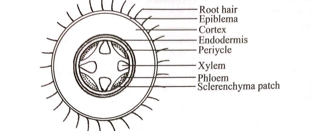
Internal structure of dicot root exhibits epidermis, cortex, endodermis, pericycle, vascular bundle, conjunctive tissue and pith.
Epidermis
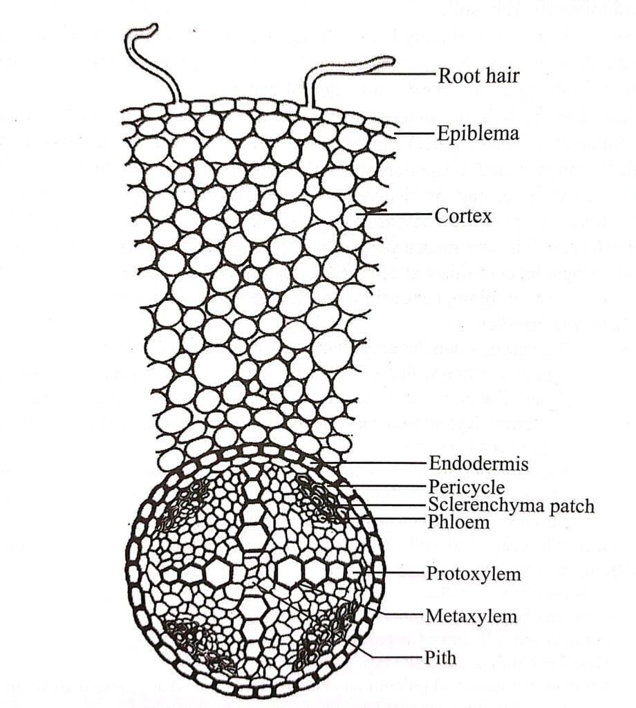
The epidermis of dicot roots consists of a single layer of thin-walled parenchymatous cells. The outer walls of these epidermal cells are not covered by a waxy substance called cuticle.
Many of these epidermal cells extend to form long, single-celled outgrowths known as root hairs. The epidermis of the root is also referred to as the epiblema or piliferous layer.
These root hairs play a crucial role in the absorption of water from the soil, aiding in the uptake of water by the plant.

Cortex
In dicot roots, the cortex is highly developed and occupies a significant portion. It consists of multiple layers of thin-walled, living parenchyma cells that have intercellular spaces.
These cells are rounded in shape and contain leucoplasts, which are responsible for converting sugar into starch grains. The last layer of the cortex is known as the endodermis, which is found universally in roots.
The primary function of the cortex is to store starch, serving as a storage reservoir for the plant.
Endodermis
The innermost layer of the cortex undergoes specialization and forms a distinct structure known as the endodermis. This layer is comprised of a single row of barrel-shaped cells that are closely packed together without any intercellular spaces.
The endodermal cells possess thickened radial walls, referred to as Casparian strips, named after Robert Caspary who first observed them in 1865. These strips are made of a water-impermeable waxy substance called suberin. The presence of Casparian strips prevents the plasmolysis of endodermal cells.
However, the endodermal cells located just outside the protoxylem, known as passage cells, do not possess such thickening.
Apart from their role in maintaining the integrity of the endodermis, these cells also store starch grains, giving rise to the term “starch sheath” for the endodermis.
The endodermis plays a crucial role in regulating the movement of fluids and air between the cortex and the vascular bundle, controlling the flow in both directions.
Pericycle
The pericycle is a layer located beneath the endodermis and is typically composed of cells that are one cell thick. These cells are thin-walled and densely packed parenchyma cells.
However, in certain species, the pericycle can be multiseriate, meaning it consists of more than one cell layer.
The cells of the pericycle have significant developmental potential. They have the ability to give rise to lateral secondary roots, which branch out from the main root.
The pericycle cells are also responsible for the formation of cork cambium, a meristematic tissue that gives rise to the cork layer in woody plants. It is important to note that the pericycle is absent in most aquatic plants and some parasitic plants.
Vascular bundle
In roots, the arrangement of vascular bundles is typically radial. The xylem and phloem components form distinct patches and are separated by non-conducting cells.
In dicotyledonous roots, the number of vascular bundles is usually limited, ranging from 2 to 6. The most common arrangement is known as tetrarch, which consists of four xylem bundles and four phloem bundles.
Xylem
In the dicotyledonous roots, the arrangement of xylem within the vascular bundles follows a specific pattern.
The protoxylem, the first-formed xylem, is positioned towards the outer edge of the bundle, adjacent to the pericycle. On the other hand, the metaxylem, which develops later, is located towards the center of the bundle.
This arrangement is known as exarch, with the younger metaxylem cells closer to the center. In some cases, the xylem bundles extend towards the pith, creating a stellate or star-shaped appearance in cross-section.
The primary function of the xylem is to transport water and minerals from the roots to the shoots of the plant. Additionally, it provides mechanical strength to the roots.
Phloem
In dicotyledonous roots, the phloem is situated between two adjacent xylem bundles. The number of xylem and phloem bundles is equal, maintaining a balanced arrangement.
The phloem is composed of sieve tubes, companion cells, and phloem parenchyma. Outside each group of phloem, there is a small patch of sclerenchyma cap.
Phloem fibers are specialized cells that provide mechanical support in some plant tissues, but they are not typically present in the primary roots of dicotyledonous plants.
Conjunctive tissue
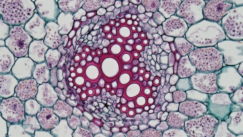
The parenchymatous tissue that lies between the xylem and phloem in the vascular bundles is referred to as conjunctive tissue. This tissue is also known as complementary tissue.
During secondary growth, the conjunctive tissue transforms into the inter-fascicular cambium, which contributes to the formation of secondary vascular tissues.
Pith
The pith, located in the center of the root, is usually not well-developed. It consists of loosely arranged parenchymatous cells that have intercellular spaces between them.
However, in certain cases, the metaxylem elements of different vascular bundles may converge and meet at the center of the root.
When this occurs, the pith region is absent as there is no distinct collection of parenchyma cells in that area.
FREQUENTLY ASKED QUESTIONS
Describe the internal structure (anatomy) of typical dicot root.
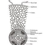
Internal structure of dicot root exhibits epidermis, cortex, endodermis, pericycle, vascular bundle, conjunctive tissue and pith.
Epidermis: Epidermis is the outermost single layer composed of thin walled, barrel shaped, compactly arranged parenchymatous cell. It consist of root hairs, responsible for absorption and helps in uptake of water.
Cortex: It is the multi-layered region inner to epidermis made up of thin walled, living parenchymatous cell containing some intracellular space. It is mainly responsible for storage of starch.
Endodermis: It is the innermost layer of cortex, composed of compactly arranged, barrel shaped cells. It consist of casparian stripes and passage cells. It is responsible for movement of fluid in or out of vascular bundle.
Pericycle: The pericycle is a layer located beneath the endodermis made of thin-walled and densely packed parenchyma cells, responsible for development of lateral roots and formation of cambium.
Vascular bundle: Vascular bundles is typically radial type wherein xylem and phloem are found in distinct patches, commonly tetrarch condition. Xylem is found alternating to phloem in exarch condition and are responsible for transport of water and minerals. Phloem are present between two adjacent xylem in the form of patches.
Conjunctive tissue: It parenchymatous tissue that lies between the xylem and phloem in the vascular bundles, forms inter-fascicular cambium during secondary growth.
Pith: The pith lies at center of the root, consist of loosely arranged parenchymatous cells, responsible for storage.

