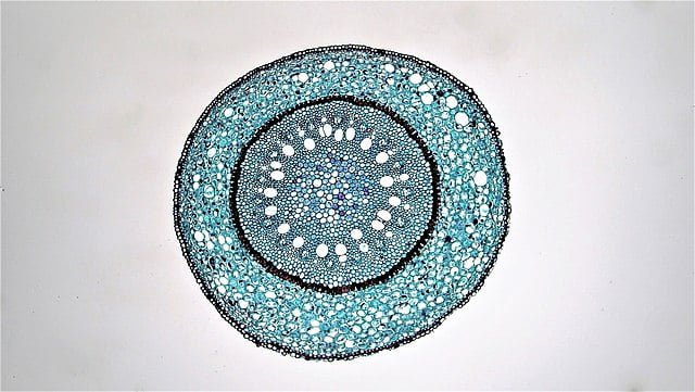
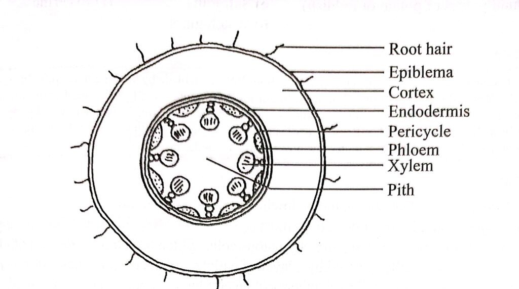
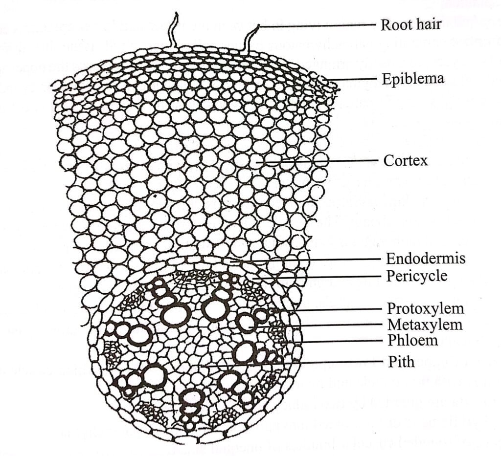
Internal structure of monocot root consist of epidermis, cortex, endodermis, pericycle, vascular bundle, conjunctive tissue and pith.
Epidermis
The outermost layer of the plant, commonly referred to as the epidermis. The term “epiblema” is not commonly used in modern plant anatomy.
Epidermis consist of thin-walled compact cells called parenchyma cells. These cells are single-celled and do not have spaces between them.
Root hairs are found in the epidermis of the roots which are tubular outgrowths that increase the surface area of the roots, facilitating the absorption of water and minerals from the soil.

Cortex
The cortex is indeed a multi-cellular layer located between the epidermis and the endodermis in plant roots. It consists of thin-walled parenchymatous cells with sufficient intercellular spaces.
The outer few layers of the cortex, in older roots, can become more compact and suberized, forming the exodermis. The exodermis acts as a protective layer, shielding the internal tissues from external injurious agents such as pathogens or toxins.
The cortical cells often contain abundant starch grains, serving as a storage reserve of food. The cortex facilitates the movement of water and minerals from the root hairs in the epidermis to the inner tissues of the root.
In older roots, the compact and suberized outer layers of the cortex form the exodermis, which acts as a protective barrier against external harmful agents.
Endodermis
The endodermis is located adjacent to the vascular tissue, separating it from the cortex. The cells of the endodermis are typically barrel-shaped and compactly arranged, lacking intercellular spaces.
In the young endodermal cells, a strip of suberin and lignin called the casparian strip is present on the radial and transverse walls. This strip acts as a waterproof barrier and controls the movement of substances through the endodermal cells.
Opposite to the protoxylem, there are thin-walled cells known as passage cells. These cells allow for the movement of fluids, such as water and dissolved nutrients, from the cortex to the xylem vessels.
The endodermis serves multiple functions. It acts as a storage layer, storing various substances within the cells. Additionally, the endodermis plays a crucial role in regulating the flow of water inward and outward from the stele (the central vascular tissue of the root).
The casparian strip and the selective transport properties of the endodermal cells enable the endodermis to control the movement of water and solutes, ensuring efficient nutrient uptake and preventing the backflow of water.

Pericycle
The pericycle is located just below the endodermis in the root. It is composed of single-layered parenchymatous cells. In certain plants the pericycle can be thick-walled and multilayered.
The pericycle forms a complete ring around the vascular tissue, including the xylem and phloem. It acts as an outermost layer of the vascular cylinder.
The pericycle has two important functions. It serves as a storage site for food reserves. These food reserves can be utilized by the plant when needed. And it plays a crucial role in initiating the development of lateral roots.
Lateral roots arise from the pericycle cells, which undergo cell divisions and differentiate into root primordia, eventually giving rise to the formation of new lateral roots.

Vascular bundle
In monocot root, vascular bundles are radial in arrangement. There are eight to twelve bundles each of xylem and phloem. Hence, the condition is described as polyarch. Exarch and polyarch vascular bundles occur in monocot root.
Xylem
The xylem tissue forms discrete strands or bundles that alternate with phloem strands. It follows an exarch arrangement, meaning that the protoxylem, the first-formed xylem elements, are located towards the periphery, while the metaxylem, the later-formed xylem elements, are towards the center.
The xylem tissue consists of vessels and xylem parenchyma. Vessels are elongated, tubular structures that allow for the efficient transport of water and minerals. Xylem parenchyma cells are associated with the vessels and provide support and storage functions.
The vessels of the protoxylem are narrow, and their walls possess annular (ring-shaped) and spiral thickenings. In contrast, the vessels of the metaxylem are broader, and their walls have reticulate (net-like) and pitted thickenings. These different types of thickenings provide strength and structural support to the xylem elements.
The primary function of the xylem tissue is the conduction of water and minerals from the roots to the stems and other aerial parts of the plant. The xylem vessels create a continuous pathway that allows for the upward movement of water and dissolved nutrients through a process known as transpiration and cohesion.

Phloem
The phloem is located as thread-like structures near the outer region of the vascular cylinder, below the pericycle. These phloem strands exhibit an exarch arrangement, where the protophloem is found towards the periphery and the metaphloem towards the center.
The phloem tissue is composed of three main types of cells: sieve elements, companion cells, and phloem parenchyma.
Its primary role is to transport nutrients, including food, to the root systems.
Conjunctive tissue
The conjunctive tissue is an area situated between the xylem and phloem strands, consisting of densely packed parenchyma cells. As roots mature, this tissue may undergo a transformation and become sclerenchymatous, characterized by the presence of hard, lignified cells.
The conjunctive tissue serves two main functions: storing food and providing mechanical support to the root structure.
Pith
The pith refers to the central region of the root, comprised of parenchyma cells that are loosely arranged and have both rounded and angular shapes. It is characterized by its large size, abundant intercellular space, and the presence of starch grains.
In certain plant species the pith may contain sclerenchyma cells with thick walls and lignified characteristics. In aquatic plants, the pith is known to have numerous air chambers.
The primary function of the pith is to serve as a storage site for food reserves.


