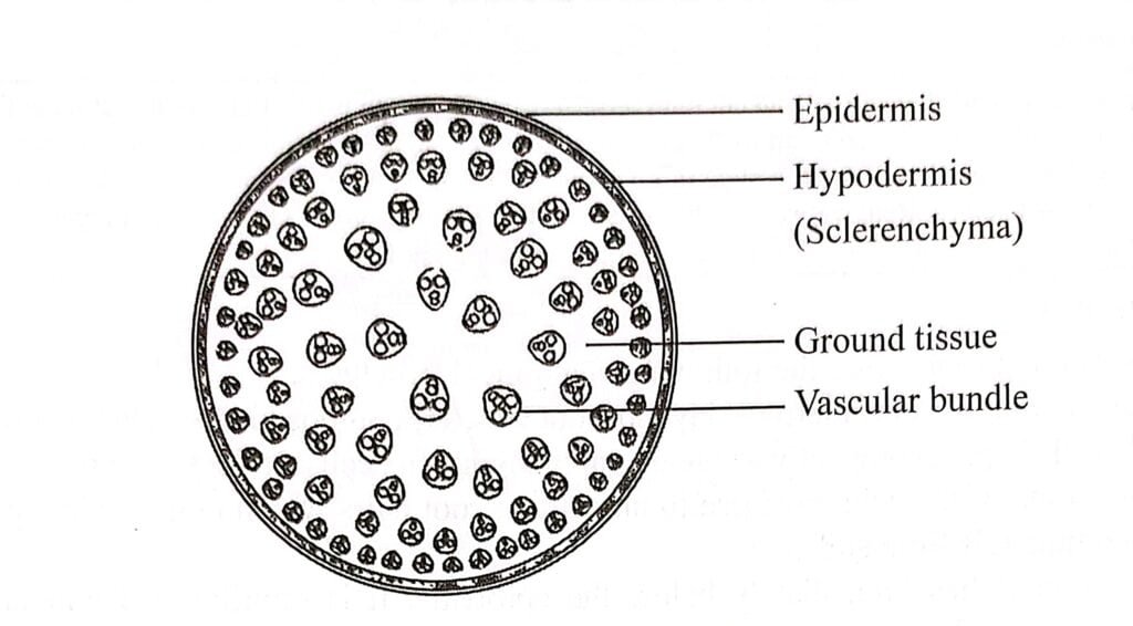
Internal structure of monocot stem possess following structures: Epidermis, Hypodermis, Ground Tissue and Vascular Strand.
Size and internal structure of monocot stem varies in different species. They consist of distinct nodes and internodes. Branches and leaves usually sessile or sub-sessile arises from nodes.
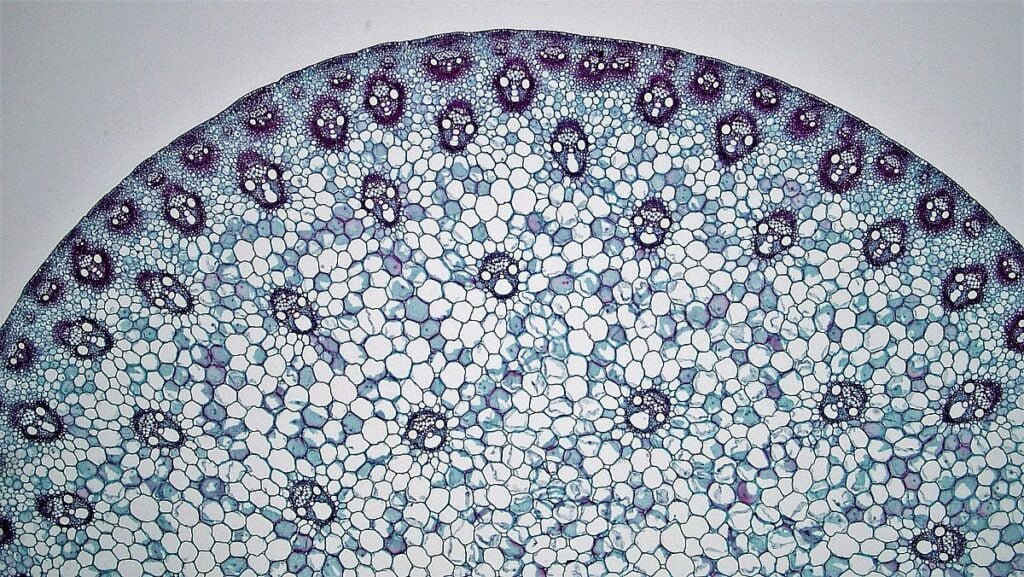
Epidermis
The stem’s outermost layer is known as the epidermis, consisting of living parenchyma cells that are transparent, elongated, and rectangular in shape, arranged tightly together.
Silica and Chitin are substances found in the outer wall of these epidermal cells. A layer called the cuticle is present on the exterior. Unlike, the epidermis of dicot stem (which consist of epidermal hairs), epidermis of monocot stem is devoid of epidermal hairs.
The cuticle and cutinized epidermal cells serve to prevent water evaporation from the stem. Additionally, silica provides rigidity. Some plants have stomata in the epidermis to facilitate the exchange of gases
Hypodermis
The hypodermis, located just beneath the epidermis, is a layer that is typically composed of a few (2-3) cell layers. These cells are thick-walled and made of lignified sclerenchyma.
Hypodermis provides strength and rigidity to the stem. The hypodermis acts as a heat screen, offering protection and insulation. Its primary functions are to enhance the structural integrity of the stem and provide support.
Ground tissue
The interior of the stem is occupied by a parenchymatous ground tissue, which lacks differentiation into cortex, endodermis, pericycle, and pith.
In maize stems, the cells are small and angular near the hypodermis, but they become large and oval in the inner region. Some outer cells are capable of synthesizing food due to the presence of chloroplasts (Chlorenchymatous cells).
The ground tissue contains abundant intercellular spaces that allow for gas exchange through the stomata present in the epidermis. Additionally, the cells store reserve food materials. The ground tissue also contains embedded vascular bundles.
Vascular bundles
The vascular system in the stem consist of vascular bundles are scattered within the ground tissue. These bundles are smaller in size, densely arranged towards the periphery, and larger with a more distant arrangement towards the center.
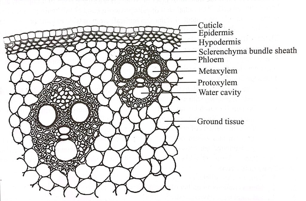
Consequently, the vascular bundles are conjoint, collateral, and closed. Each vascular bundle is surrounded by a sclerenchyma sheath called the bundle sheath, which is more developed on the outer and inner sides.
In some outer vascular bundles, the hypodermis and bundle sheaths merge together. The vascular bundles have an oval or rounded shape and contain both phloem and xylem tissues. The phloem is located towards the outer side, while the xylem is positioned on the inner side.
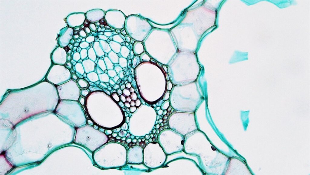
Xylem
The Y-shaped vascular system is present in the stem. The xylem tissue is endarch, meaning that the protoxylem is located towards the center of the stem. It consists of vessels, tracheids, xylem parenchyma, and a few xylem fibers.
The metaxylem usually contains two large oval or rounded vessels situated at the upper two angles of the xylem region. The protoxylem, on the other hand, consists of a small number (2-3) of small vessels located at the lower angle of the xylem.
During the rapid growth of the stem, some of the protoxylem vessels and xylem parenchyma cells dissolve or separate, creating a cavity called the water cavity, lysigenous cavity.
The tracheids and vessels play a role in sap conduction and also provide mechanical support. Lysigenous cavity generally functions as a water storage area.
Phloem
The phloem tissue is composed of sieve tubes, companion cells, and a small number of phloem fibers. The phloem can be further divided into protophloem and inner metaphloem.
The protophloem tends to get compressed or crushed during the later stages of development.
Phloem parenchyma is not present. The sieve tubes are responsible for conducting organic food substances.

