Mitochondria are tiny, granular or filament-like autonomous cell organelles located in the cytoplasm of cells of aerobic eukaryotes, mainly responsible for production of energy in the form of ATP, regulation of cell cycle, cell signaling etc.
They are absent in prokaryotic cell and anaerobic eukaryotes.
These well-defined organelles play essential roles in various cellular metabolic functions, often earning them the title “powerhouses of the cell.” Mitochondria produce the majority of the cell’s adenosine triphosphate (ATP), which is used as a primary energy source.
They contain a range of enzymes and coenzymes that catalyze energy transformations within the cell. Additionally, mitochondrion house specific DNA for cytoplasmic inheritance and ribosomes for protein synthesis.
Energy is obtained through the breakdown of carbohydrates, amino acids, and fatty acids, which is then used to form ATP—commonly known as the cell’s “energy currency”—via oxidative phosphorylation.
Beyond providing energy, mitochondrion also play roles in cell signaling, differentiation, and programmed cell death, while helping regulate the cell cycle and growth.

A Short Historical Perspective
- In 1880, Rudolf Kölliker was the first to observe mitochondrial granules in the striated muscle cells of insects.
- In 1890, Richard Altmann recognized these structures as cell organelles and named them “bioblasts.”
- In 1898, Carl Benda coined the term “mitochondria”.
- In 1900, Leonor Michaelis found that Janus green could be used as a supravital stain to visualize mitochondria.
- In 1904, Friedrich Meves made the first recorded observation of mitochondria in plants, specifically in the cells of the white waterlily, Nymphaea alba.
- In 1908, Regaud, concluded that mitochondrion consist some proteins and lipids. And he along with Meves, revealed that it contains genes.
- In 1912, Kingsbury proposed that mitochondrion as the site for internal respiration.
- In 1920s, biochemist Otto Warburg discovered that oxidative reactions occur within specific cell regions in most tissues.
- In 1934, Bensley and Hoerr, made first attempt to isolate mitochondria by cell fraction method.
- In 1948, Hogeboom and his co-workers isolated morphologically well-preserved mitochondria.
- In the early 1950s, Palade and Sjöstrand illustrated double-membrane structure of mitochondrion is made up of an inner and outer membrane.
- In 1957, Philip Seekevitz, termed mitochondria as “power house of cell”.
- In 1960, Racker and his colleagues isolated enzyme ATP synthase form mitochondrion.
Morphology of Mitochondria
- Mitochondrion can appear as filaments or small granules, sometimes taking on a rod-like shape known as chondriosomes. These structures can grow or cluster into larger spherical bodies called chondriospheres.
- Location-wise, mitochondria are generally dispersed throughout the cytoplasm, with the ability to move independently. In some cells, they can travel freely, delivering ATP to areas requiring energy, while in others, they are permanently situated near high-energy-demand regions. During cell division, mitochondria often gather around the spindle apparatus.
- The number of mitochondria varies significantly between different cell types and species. Some algae and certain protozoa have only a single mitochondrion, while the amoeba Chaos chaos may contain up to 50,000. In rat liver cells, the count ranges between 1,000 and 1,600, and in some oocytes, it can reach up to 300,000 mitochondria.
- The typical mitochondrion size is around 0.5-1.0 µm in diameter and 2-8 µm in length. In mammalian pancreatic exocrine cells, it can be as long as 10 µm, while in the oocytes of the amphibian Rana pipiens, they may reach 20-40 µm. Yeast cells contain some of the smallest mitochondria.
Ultra Structure of Mitochondria
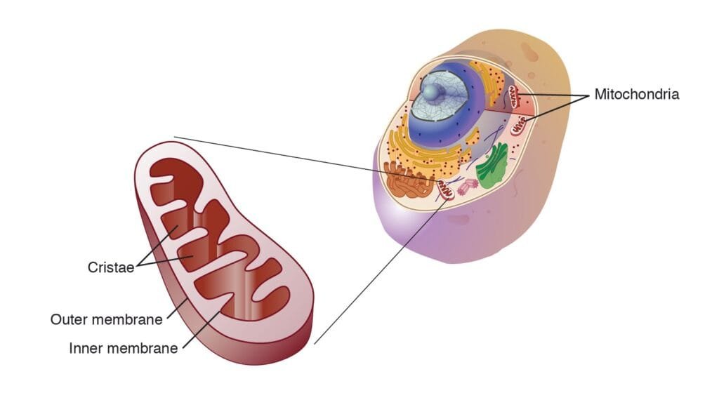
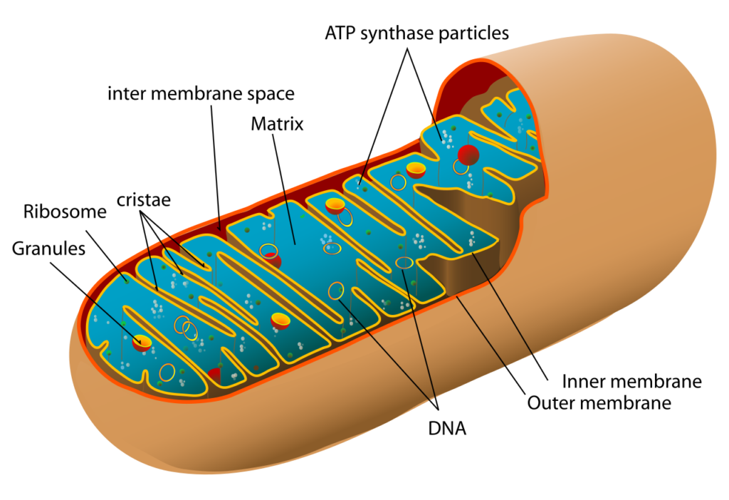
Mitochondria are double-membraned organelles, with each mitochondrion surrounded by two highly specialized membranes. The outer membrane, about 6 nm thick, encases the organelle, and within it lies an inner membrane separated by a 6-8 nm space.
While the outer membrane is smooth and continuous, the inner membrane extends into the mitochondrial cavity, forming complex folds called crests. These folds, known as cristae, protrude into the matrix.
The two membranes, distinct in chemical and functional properties, create two separate compartments within the mitochondrion: the matrix and the intermembrane space.
This double-membrane structure divides the mitochondrion into five distinct parts.
- Outer membrane
- Intermembrane space
- Inner membrane
- Cristae
- Matrix

Outer Membrane
- The outer membrane, which encloses the organelle, is approximately 60 to 75 Å thick and consists of a simple phospholipid bilayer, made up of 50% lipids and 50% proteins by weight.
- This membrane resembles the outer membrane found in the cell walls of certain bacteria. It is highly permeable due to the presence of numerous integral proteins, especially porins.
- The outer membrane also contains enzymes involved in various functions, including fatty acid elongation, mitochondrial lipid synthesis, epinephrine oxidation, and tryptophan degradation.
- Mitochondrion that have lost their outer membrane are referred to as mitoplasts.
Intermembrane Space
- The outer chamber, or intermembrane space, lies between the outer and inner membranes, typically spanning 60–75 Å and filled with a watery fluid containing a few enzymes.
- Also called the peri-mitochondrial space, it can expand if isolated mitochondria are placed in a sucrose solution.
- This space plays a critical role in the primary function of mitochondria—oxidative phosphorylation.
- It also houses enzymes that utilize ATP to phosphorylate other nucleotides.
Inner Membrane
- The inner membrane of the mitochondrion is impermeable to most ions and small charged molecules, acting as a functional barrier that restricts free movement between the cytosol and the matrix.
- Composed of over 100 distinct polypeptides, it has a high protein-to-lipid ratio (80:20).
- This membrane is the main site for ATP synthesis and is folded into finger-like projections known as cristae, which increase the membrane’s surface area.
- The number and shape of these cristae can vary significantly; higher ATP demands result in a greater number of cristae.
- The inner membrane is complex, containing electron transport chain complexes, ATP synthase, and transport proteins.
- The primary roles of proteins in the inner membrane include; oxidative phosphorylation, ATP production, regulation of protein transport, protein import, mitochondrial fusion and fission
- The inner membrane separates the mitochondrion into two compartments: the outer chamber, which is located between the two membranes and within the core of the cristae, and the inner chamber, which is filled with a relatively dense protein-rich substance known as the mitochondrial matrix.
Cristae
- Typically, the cristae are incomplete septa or ridges that maintain the continuity of the inner chamber, ensuring that the matrix remains continuous throughout the mitochondrion.
- The inner membrane is organized into numerous cristae, which increase the surface area of the membrane and improve its capacity to generate ATP.
- The cristae and the inner boundary membranes are connected by junctions referred to as cristae junctions.
- The folds of the cristae membranes are adorned with small, round protein complexes known as F1 particles or oxysomes, which are the sites of ATP synthesis driven by a proton gradient.
F1 Particles
- The inner membrane in the cristae is covered with particles measuring 8.5 nm, each connected to the membrane by a stem.
- These particles, known as elementary particles (F1 or F0-F1 particles), are evenly spaced at intervals of 10 nm on the inner membrane’s surface.
- A single mitochondrion may contain between 10^4 and 10^5 elementary particles, which correspond to a specialized ATP synthase that plays a crucial role in the coupling of oxidation and phosphorylation.
- Electron micrographs show that the ATP synthase, or F1 particle, attached to the inner membrane consists of two main components: the F1 head piece, which contains five different subunits—alpha (α), beta (β), gamma (γ), delta (δ), and epsilon (ε)—and the F0 base piece, which is embedded in the membrane.
- The subunit of F0 extends into the head piece to form the stalk (stem).
Matrix
- The matrix is the space enclosed by the inner membrane of the mitochondrion and contains approximately two-thirds of the organelle’s total protein.
- It plays a crucial role in ATP production with the assistance of ATP synthase found in the inner membrane. The matrix is a concentrated mixture of hundreds of enzymes necessary for expressing mitochondrial genes, along with insoluble inorganic salts, specialized ribosomes, tRNA, and multiple copies of the mitochondrial DNA genome.
- Additionally, it houses the enzymes needed for the oxidation of pyruvate and fatty acids, as well as for the citric acid cycle.
- Mitochondrial DNA is responsible for cellular respiration, and it possess their own genetic material, enabling them to produce their own RNAs and proteins.
- This genetic independence makes the mitochondrion semi-autonomous.
Function of Mitochondria
Mitochondria provide almost all the biological energy needed by cells. The ATP produced during cellular respiration accumulates within it.
Mitochondrial genes affect specific hereditary traits, such as male sterility in maize, and organisms usually receive their mitochondria from their mothers, a phenomenon known as maternal inheritance.
Mitochondrion function as small biochemical factories where food substances or respiratory substrates are fully oxidized into carbon dioxide and water. Due to their role in ATP production, mitochondria are often referred to as the powerhouses of the cell.
Some key functions of mitochondria include:
- ATP Production: The primary function of mitochondria is to generate energy through oxidative phosphorylation. They act as energy-transducing organelles where the major degradation products of cellular metabolism are transformed into chemical energy (ATP) for various cellular activities. The energy transformation processes involve three coordinated steps: the Krebs cycle, the electron transport chain, and ATP phosphorylation.
- Fatty Acid Synthesis: The matrix, or inner chamber contains enzymes responsible for fatty acid synthesis. Within the matrix, each fatty acid molecule undergoes complete breakdown through a series of reactions known as β-oxidation, resulting in acetyl-CoA, which then enters the Krebs cycle for further process.
- Synthesis of Biochemicals: It supply essential intermediates for the production of various biochemicals, including chlorophyll, cytochromes, pyrimidines, steroids, and alkaloids. In addition to ATP production, it also engage in certain biosynthetic or anabolic processes.
- DNA and Protein Synthesis Machinery: Mitochondrion contain their own DNA and the necessary machinery for protein synthesis, allowing them to produce specific proteins independently. These include subunits of ATP synthase, components of reductase, and three out of seven subunits found in cytochrome oxidase.
- Haemo Synthesis: The synthesis of haem, which is essential for myoglobin and haemoglobin, begins with a mitochondrial reaction facilitated by the enzyme delta-aminolevulinic acid synthetase.
- Cholesterol Conversion: Some initial steps in converting cholesterol to steroid hormones in the adrenal cortex are also catalyzed by enzymes located in the mitochondria.
- Storage Functions: It can also involve on storage roles; for instance, the mitochondria in ova store large quantities of yolk proteins and convert them into yolk platelets.
- Calcium Ion Regulation: It help cells maintain appropriate calcium ion concentrations within various compartments. They can store and release calcium as needed.
- Blood and Hormone Production: They assist in the synthesis of certain components of blood and hormones such as testosterone and estrogen.
- Ammonia Detoxification: The mitochondria in liver cells contain enzymes that are responsible for detoxifying ammonia.
- Role in Apoptosis: They are crucial in the process of apoptosis, or programmed cell death. Dysfunction in mitochondria can lead to abnormal cell death, which may impact organ function.
Summary
Mitochondria are tiny, granular or filament-like organelles located in the cytoplasm of aerobic cells in higher animals, plants, and some microorganisms, including protozoa, algae, and fungi. They are absent in prokaryotic cells and anaerobic eukaryotes.
They produce the majority of the cell’s adenosine triphosphate (ATP), which is used as a primary energy source. Due to their role in ATP production, mitochondria are often referred to as the powerhouses of the cell.
Found to be dispersed through out the cytopalsm, mostly concentrated to high energy demanding regions. They vary in number form single to hundreds of thousad dependig upon the types of organisms or types of cell in an organism.
It is composed of double layered membranous structure outer and inner membrane, consisting intermembranous space in between them. Matrix is present enclosed inside inner membrane which corresponds to cytoplasm consist of mixture of hundreds of enzymes necessary for expressing mitochondrial genes, along with insoluble inorganic salts, specialized mitochondrial ribosomes, tRNA, and multiple copies of the mitochondrial DNA genome. Inner membrane is organized into numerous cristae which are covered by F1 particles which play important role in oxidative phosphorylation.
Primary function of mitochondria is the synthesis of ATP, fatty acids, intermediate complexes as cytochromes, pyrimidines, steroids etc. Beside that other function of mitochondria is to help in the storage, regulation of Calcium ion concentration, production of hormones, detoxification of ammonia and involve in programmed cell death.


